If you're in need of a cell culture plate that offers the following benefits:
1.Allows direct absorbance measurement in a microplate reader
2.Enables easy observation of cell growth under a microscope
3.Facilitates the cultivation of luminescent cells while preventing interference between wells
Look no further than NEST Black/White Clear Bottom Cell Culture Plates.

NEST Black/White Clear Bottom Cell Culture Plates are made of non-autofluorescent materials and undergo special surface treatment to significantly enhance their protein binding capability. Unlike regular black and white culture plates, NEST plates have well bottoms made of high-transparency PS material, which allows for excellent light transmission and enables use in high-magnification microscopes and microplate readers.
The well walls are made of light-shielded PS material, which prevents cross-well interference seen in fully transparent plates. Additionally, this design allows for microscope observation, a limitation of fully black or white enzyme-linked immunosorbent assay (ELISA) plates.
Black Clear Bottom Cell Culture Plate
The black clear bottom cell culture plate is designed to effectively minimize background fluorescence. With its superior light absorption capabilities, it absorbs background fluorescence signals, reducing interference and improving the signal-to-noise ratio. This enhances the sensitivity and accuracy of fluorescence measurements. The black opaque well walls also prevent cross-interference between wells.
Application: Widely utilized for cell immunofluorescence detection under intense light conditions.

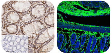
Cell immunofluorescence detection
Cell immunofluorescence detection is a technique based on the reaction of antigens and antibodies. In this method, known antigens or antibodies are labeled with fluorescent tags to create fluorescent markers. These fluorescently labeled antibodies (or antigens) act as molecular probes to identify specific antigens (or antibodies) within cells or tissues. The formation of antigen-antibody complexes in cells or tissues results in the presence of fluorophores. When observed under a fluorescence microscope, these fluorophores emit bright fluorescence (in hues of yellow-green or orange-red) upon excitation by light, enabling the visualization of cells or tissues with fluorescence. This approach aids in determining the characteristics and location of antigens or antibodies, as well as quantifying their levels using quantitative methods.

White Clear Bottom Cell Culture Plate
NEST White Clear Bottom Cell Culture Plate, when used as a background, provides a uniform and stable background light. The reflective and scattering properties of the white plate help reduce the absorption of chemiluminescent signals. Additionally, it illuminates the entire test area, resulting in a more uniform distribution of chemiluminescent signals. This is particularly beneficial for detecting samples with low luminescence, as using a white plate can significantly improve the signal-to-noise ratio.
Application: These plates are commonly utilized in immunohistochemistry testing with low light, including general chemiluminescence and substrate coloration experiments.

Immunohistochemistry (IHC) is a laboratory technique that depends on the reactions of antigens and antibodies. By using chemical reactions to visualize labeled antibodies, IHC allows for accurate identification, qualitative evaluation, and quantitative measurement of specific antigens or antibodies in tissues and cells. Providing a detailed assessment of changes in protein expression at a microscopic level during disease progression, IHC holds significant value in both pathological diagnosis and research. It enables the examination of colored cells or tissues under a standard optical microscope and facilitates the determination of the total amount of colored substances using an enzyme immunoassay instrument.
Buying Guide